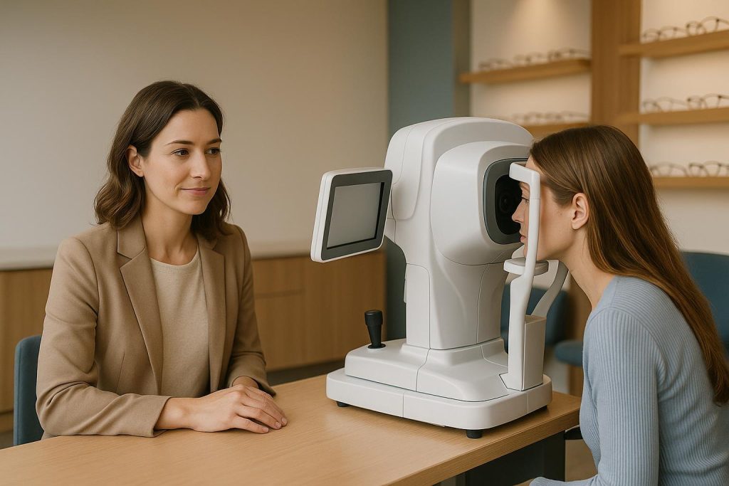
How Retinal Detachment Is Diagnosed and Why Fast Care Matters
A retinal detachment is diagnosed through a prompt, comprehensive eye examination that lets an optometrist or ophthalmologist look closely at the retina at the back of the eye. The retina is the light-sensitive tissue that sends visual information to the brain. When it pulls away from the layer beneath it, its blood and nutrient supply can be affected. This can lead to permanent vision loss if it is not treated quickly.
You cannot reliably test for retinal detachment at home. Symptoms can be an important warning, but only an in-person eye examination can confirm whether there is a retinal tear, retinal hole, detachment, or another cause of sudden visual changes. If you notice new flashes, a sudden shower of floaters, a dark curtain or shadow in your vision, or sudden loss of vision, seek urgent eye care the same day. Emergency eye exams can help assess urgent symptoms and arrange timely referral when specialist treatment is needed.
The main test: a dilated retinal examination
The most important test for suspected retinal detachment is a dilated eye exam. The eye doctor places dilating drops in your eyes to widen the pupils. This provides a much broader and clearer view of the retina, including the peripheral retina where tears often begin.
Once your pupils are dilated, the doctor uses lights and specialized lenses to examine the retina for:
- Retinal tears or holes: Small openings that may allow fluid to pass underneath the retina.
- Retinal detachment: Areas where the retina has lifted away from the tissue beneath it.
- Vitreous changes: Changes in the gel inside the eye that may pull on the retina.
- Bleeding or inflammation: Findings that may signal a tear or another eye condition.
- Other retinal concerns: Conditions such as diabetic eye disease or age-related changes that may affect retinal health.
Dilation can make your vision blurry and more sensitive to light for several hours. It is often wise to bring sunglasses and arrange a ride if you do not feel comfortable driving afterward. Although dilation may feel inconvenient, it is a valuable part of a thorough retinal health assessment.
Widefield retinal imaging and Optomap imaging
Modern retinal imaging can also play an important role in detecting and documenting retinal concerns. Widefield imaging, sometimes called Optomap imaging, captures a broad digital view of the retina. These images can help the eye care team identify changes, compare findings over time, and explain what they see in a clear way.
Retinal photographs are useful for preventative care, but they do not always replace dilation when symptoms suggest a retinal tear or detachment. Some far-peripheral areas of the retina may still need a direct dilated examination. Your optometrist will decide which tests are appropriate based on your symptoms, health history, and examination findings.
Other tests that may be used
Depending on what the doctor sees, additional testing may be needed to assess the retina and plan next steps. These tests are selected for a specific reason; not every patient requires every test.
- Optical coherence tomography (OCT): OCT uses light to create detailed cross-section images of the retina. It is especially helpful for assessing the central retina, called the macula, and for identifying fluid or subtle changes in retinal layers.
- Visual field testing: This test checks for missing areas in your side vision. A retinal detachment can create a field defect that may feel like a curtain, veil, or shadow.
- Eye ultrasound: If the view of the retina is blocked by bleeding, a dense cataract, or another issue, ultrasound may help detect a detachment behind the obstruction.
- Eye pressure and pupil testing: These are often part of an urgent eye assessment. They do not diagnose retinal detachment on their own, but they provide useful information about overall eye health.
Symptoms that need same-day assessment
Retinal detachment is not usually painful. That is why it is important not to wait for pain before seeking help. Many symptoms are caused by less serious changes, but it is impossible to know the cause without an examination. Contact an eye care professional promptly if you experience any of the following:
- New flashes of light, especially at the edge of your vision or in a dark room.
- A sudden increase in floaters, such as dots, specks, cobwebs, or squiggly lines.
- A shower of tiny dark spots or new cloud-like debris in your vision.
- A gray shadow, curtain, or veil that seems to move across part of your visual field.
- Sudden blurred, distorted, or reduced vision in one eye.
- Loss of side vision or a missing area of vision.
If these symptoms start suddenly, worsen, or occur after an eye injury, do not wait for a routine appointment. A prompt assessment is important because early treatment of a retinal tear may help prevent a larger detachment. If you have severe vision loss or cannot access urgent eye care promptly, go to the nearest emergency department.
What happens if retinal detachment is suspected?
If an optometrist suspects a retinal tear or detachment, the priority is to make sure you reach the right level of care quickly. This commonly involves urgent referral to a retinal specialist or ophthalmologist. The specialist confirms the diagnosis and determines whether treatment is needed immediately.
Treatment depends on the type, size, and location of the retinal problem. A retinal tear may sometimes be treated with laser therapy or a freezing treatment called cryotherapy to seal the retina around the tear. A true retinal detachment may require surgery, such as a gas bubble procedure, scleral buckle, or vitrectomy. The exact treatment is determined by the ophthalmologist.
Not every floater or flash means retinal detachment, and not every retinal change requires surgery. However, it is always safer to have sudden symptoms checked promptly than to assume they will pass. Early diagnosis gives the treating team the best chance to protect vision.
Who has a higher risk of retinal detachment?
Anyone can develop a retinal detachment, but some people have a higher risk. Knowing your risk can help you take changes in vision seriously and keep up with recommended eye examinations.
- People with high myopia, also called significant nearsightedness.
- People who have had eye surgery, including cataract surgery.
- Those with a previous retinal tear or detachment in either eye.
- People with a family history of retinal detachment.
- Anyone who has had a serious eye injury.
- People with certain eye conditions that affect the retina or vitreous.
- Adults experiencing age-related changes in the vitreous gel, including posterior vitreous detachment.
Having one or more risk factors does not mean you will have a retinal detachment. It does mean regular eye health monitoring and fast attention to new symptoms are especially important. Comprehensive eye exams help establish a record of your retinal health and identify concerns that may need follow-up.
Can a routine eye exam find retinal problems before symptoms begin?
Often, yes. A routine eye exam can reveal retinal thinning, holes, tears, or other changes before you notice a major change in vision. This is one reason preventative care matters, even when your glasses prescription seems stable. The examination is not only about seeing clearly; it also checks the health of structures you cannot assess on your own.
Adults can learn more about scheduling a preventative visit through adult eye exams. For older adults, regular monitoring is also valuable because several eye conditions become more common with age; see the information on senior eye exams.
What to do while waiting for urgent care
If you are experiencing symptoms that may be linked to retinal detachment, call for urgent guidance right away. Avoid driving yourself if your vision is impaired or your eyes have been dilated. Do not rub or press on the eye, especially after an injury. Make a note of when symptoms started, which eye is affected, and whether the shadow or vision loss is spreading. This information can help the examining doctor understand the situation.
Most importantly, do not try to diagnose the problem through online images or vision tests. A retinal detachment can only be assessed accurately with the proper equipment and an in-person examination. Prompt, personalized care is the safest response to sudden changes in vision.
When to book an exam
Book a routine comprehensive eye exam if you are due for one, have a high-risk history, or have noticed gradual changes in vision. Seek same-day care for new flashes, floaters, a curtain-like shadow, or sudden vision loss. Retinal detachment is a time-sensitive condition, but fast assessment and referral can make a meaningful difference in protecting your sight.